What is a pregnancy ultrasound?
Ultrasounds during pregnancy: An ultrasound scan is a useful tool not just for pregnant women but parents-to-be in general. It allows you to check up on your baby and the different stages it’s going through during gestation. An ultrasound will help your doctor or nurse practitioner check on the health and development of your baby during its entire gestation period.
Ultrasounds during pregnancy can be quite fantastic, in fact, they are the first time you get to see your own baby! However, depending on when it is done and your baby’s position at the time, you may be able to watch them move their tiny limbs. Whether you are curious about the gender of the child or if you just want privacy for yourself, let your ultrasound surgeon know-how (s)he should address this for you!
What is the normal schedule for ultrasounds during pregnancy?
- Typically, two ultrasounds are needed for a healthy pregnancy to assess the health of a child: one near the 11th week and another at about the 18th week.
- When abnormalities occur in your body, the consequences are serious. So, there’s not only one kind of ultrasound you might need to confirm what is happening with your child or yourself.
- In the final month of pregnancy, especially if the mother happens to be high-risk or the baby seems to be on its way towards becoming what is called “big for gestational age” — often due to a mother’s weight, age, or health history – it may be recommended that both patients go in for weekly ultrasounds.
What are some reasons for having an ultrasound?
Ultrasounds during pregnancy are used to perform a few different procedures, some of which include:
- To make sure that you’re pregnant.
- determine your baby’s age and growth inside you. This allows the doctor to write down the due date.
- Ultrasound is a great way for us to check on your baby’s growth and development. It can help us see how your baby has grown, and even watch the first flutter of movements!
- determine whether you are carrying multiples such as twins, triplets or more.
- When you’re expecting a baby, one of the most nerve-wracking times is when there are still some weeks to go before your due date. Your provider will likely use ultrasound technology to check in on your baby and determine whether he or she is in the head-first position or if it’s facing another way that could pose a risk for complications during birth.
- Your provider uses ultrasound to examine the internal organs responsible for reproduction and development, especially the ovaries which contain eggs, and the uterus where the baby develops.
Is it safe to have multiple ultrasounds during pregnancy?
Repeated ultrasounds are safe for the fetus. Several studies have confirmed that there is no lasting damage to the baby due to multiple ultrasounds. It was previously thought that a baby exposed to multiple ultrasounds during pregnancy would end up smaller as an infant or developing normally, but new University of Michigan research finds that repeated ultrasound exposure is associated with slowed fetal growth in late-term pregnancies.
How to prepare for an ultrasound during pregnancy?
During an ultrasound earlier in your hopes of becoming a parent, you will have to drink two to three glasses of water one hour before your scheduled ultrasound. You should not urinate prior to arriving, so it’s important that you drink water beforehand in order to arrive at your appointment with a full bladder. It’s important for the technician to get a clear image of the child without interruption or anything blocking their view.
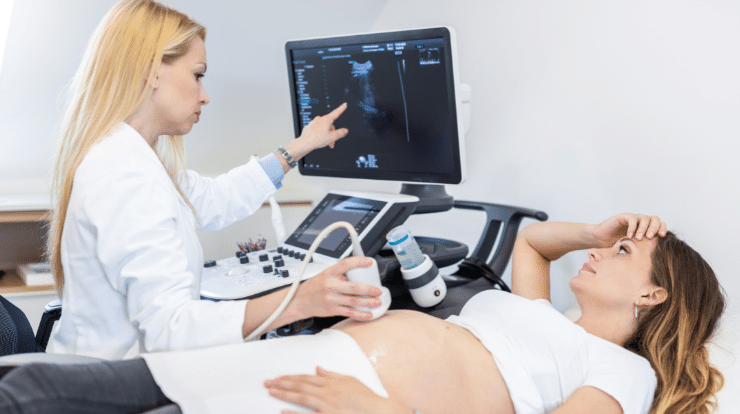
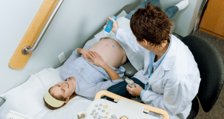
What happens during an ultrasound?
During an ultrasound, you place your feet in special clips or straps and lie on a special table. Your abdomen and pelvic area are then cleaned with a water-based gel which should not leave marks on your skin or clothes and is applied to help with the transmission of sound waves. After cleaning, the technician uses a small wand called a transducer and holds it against your skin for quick scans of the area.
A conductivity gel is also applied at this point to aid communication between the sensor and other electronic equipment located around the room. The scan takes only a few minutes – allowing time for patient responses (such as drastic movements) to be considered if necessary, at which point proper images can be captured before wiping off any remaining gel (if there are any).
Types of ultrasounds during pregnancy:
When it comes to more advanced ultrasound techniques, they’re actually more accurate and provide more information than the traditionally used ones. If the doctor detects problems during your traditional ultrasound, they’ll usually use a more advanced technique to acquire even greater information which will give them all of the necessary results they need to diagnose any potential problems.
1). Transvaginal ultrasound:
A transvaginal ultrasound scan is done to capture a clearer image of the baby. This method comes into play as early as mid-pregnancy when it might be more difficult to get clear, distinct photos of the baby. For this test, a small ultrasound probe is inserted directly into your vaginal canal to gather images of your growing bundle of joy!
2). 3-D ultrasounds during pregnancy:
A 3D ultrasound reveals each and every detail of the fetus; even its heartbeat can be heard! The 3D ultrasound is newer and more advanced compared to conventional sonography. It provides a view that shows width, length and height which allows your doctor to determine the actual age of the fetus in comparison to what was believed at the time you were born. This test helps your doctor diagnose any problems that occur during pregnancy. Also, unlike a standard ultrasound, it takes special schooling for adjudicators – so it may not be readily available.
3). 4-D ultrasounds during pregnancy:
A 4-D ultrasound can be referred to as a dynamic 3-D ultrasound. This kind of ultrasound creates a video of the baby and its movements from multiple angles rather than showing the images as regular 2-D ultrasounds do. It is used to get better views of the fetus and its facial features. As you might have already seen, this type of ultrasound is performed in a similar way to other ultrasounds: only with special equipment.
4). Fetal echocardiography:
Fetal echocardiography is similar to traditional ultrasound, except it will usually last longer. During this medical procedure, the heart’s size, shape, and structure are being analyzed in detail. An expert can see how the baby’s heart functions, which may be helpful in diagnosing congenital heart defects.